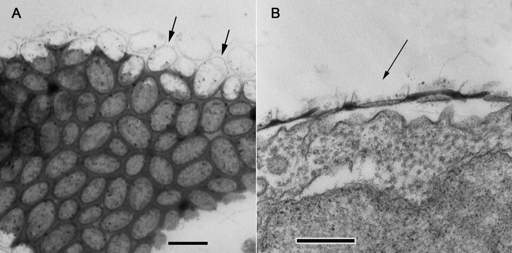
Fig. 3.
Higher magnification images of ultrathin sections through the electron dense organic coat and surface scales of the amoeba. A. Approximately tangential section (slightly oblique to the coat surface) showing the organization of the scales, particularly the oval outlines of the scale perimeters (arrows). B. Transverse section through the vertical thickness of the organic coat showing a more detailed profile of one of the scales (arrow) in vertical profile. Scale bars = 200 nm.

