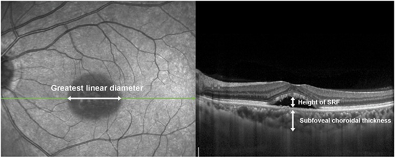
Figure 1.
Measurements of the greatest linear diameter, subfoveal choroidal thickness, and height of SRF. On SD-OCT, the greatest linear diameter of lesion boundaries, as basal area of detached retina, was measured (left). Also, the height of SRF was defined as the greatest distance from the retinal pigment epithelium (RPE) to the border of the detached neurosensory retina, within an area from 3 mm nasal and 3 mm temporal from the center of the fovea (right). Subfoveal choroidal thickness was measured from the RPE to the inner surface of the sclera within an area at the center of the fovea, on enhanced depth imaging optical coherence tomography (EDI-OCT) (right).

