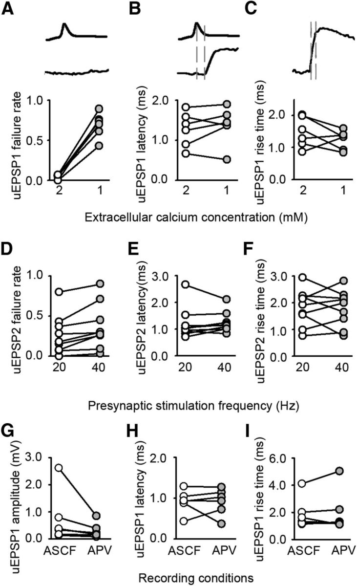
Figure 3.

Altered timing of neurotransmission in deprived cortex is independent of release probability and NMDA receptor activation. A–C, Effect of decreasing the extracellular calcium concentration on uEPSP1. A, Failure rate (2 mm 0.04 ± 0.01; 1 mm 0.69 ± 0.06; n = 6, p < 0.001, paired t test). B, Latency (2 mm 1.3 ± 0.2 ms; 1 mm 1.4 ± 0.2 ms; n = 6, p = 0.380, paired t test), C, Rise time (2 mm 1.5 ± 0.2 ms; 1 mm 1.2 ± 0.1 ms; n = 6, p = 0.255, paired t test). D–F, Effect of increasing the frequency of presynaptic firing from 20 to 40 Hz on uEPSP2. D, Failure rate (20 Hz = 0.26 ± 0.08, 40 Hz = 0.37 ± 0.09, p = 0.007, paired t test, n = 9). E, Latency (20 Hz = 1.2 ± 0.2 ms, 40 Hz = 1.2 ± 0.1 ms, p = 0.651, paired t test, n = 9). F, Rise time (20 Hz = 1.8 ± 0.2 ms, 40 Hz = 1.8 ± 0.2 ms, p = 0.861, paired t test, n = 9). G–I, Effect of blocking NMDA receptors on uEPSP1. G, Amplitude (before APV 0.36 [0.16–1.3] mV, after APV 0.19 [0.13–0.51] mV, n = 6 connections; p = 0.031, Wilcoxon signed rank test). H, Latency (before APV 0.92 ± 0.11 ms, after APV 0.91 ± 0.13 ms, n = 6 connections; p = 0.949, paired t test). I, Rise time (before APV 1.90 ± 0.46 ms, after APV 2.02 ± 0.63 ms, n = 6 connections; p = 0.574, paired t test).
