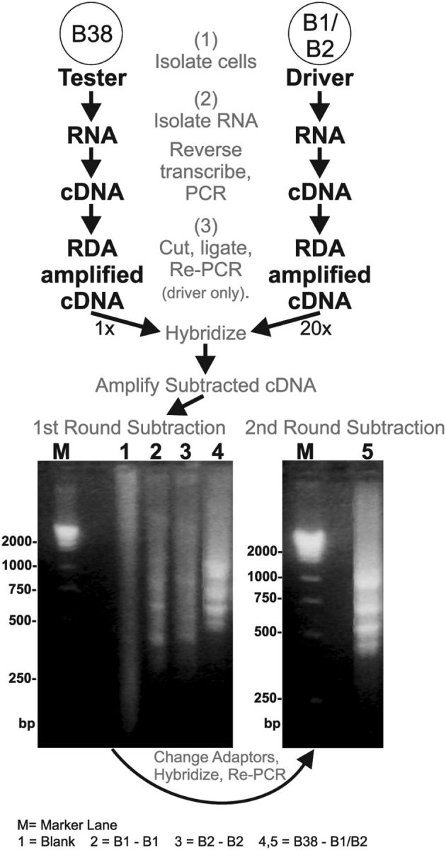
Figure 1.

RDA procedure used to identify ApVGLUT. Illustrated is the RDA between a glutamatergic neuron, B38, and B1/B2 neurons. The results of the subtractions performed are shown in the gels at the bottom. Bottom left, The results of the first subtraction performed. Lane 1 is a blank subtraction. Lanes 2 and 3 are additional control subtractions using the B1 or B2 neurons subtracted against themselves, respectively. These lanes only show a weak smear with some faint bands. Lane 4 is a B38 that was subtracted against neurons B1 and B2. This lane shows some intense bands that do not appear in the control subtractions. M, Marker lane showing cDNA masses in base pairs. Bottom right, Lane 5 shows the results of a second round of subtraction. Among several clones, one difference clone obtained from B38 was sequenced to reveal an open reading frame that coded for a protein named ApVGLUT.
