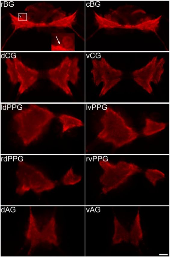
Figure 6.

Immunocytochemistry of ApVGLUT in the Aplysia CNS. Immunostaining reveals that the majority of the ApVGLUT protein is located in the neuropil and intense immunostaining of the neuropil is observed in all ganglia. Based on its location in the rostral buccal ganglion, a putative B38 (arrow) was identified. The antibody against ApVGLUT was iCT. rBG, rostral buccal ganglion; cBG, caudal buccal ganglion; dCG, dorsal cerebral ganglion; vCG, ventral cerebral ganglion; ldPPG, left dorsal pleural and pedal ganglia; lvPPG, left ventral pleural and pedal ganglia; rdPPG, right dorsal pleural and pedal ganglia; rvPPG, right ventral pleural and pedal ganglia; dAG, dorsal abdominal ganglion; vAG, ventral abdominal ganglion; Scale bar, 200 μm.
