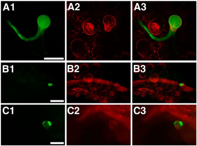
Figure 8.
B64 is ApVGLUT positive. A1–C1, B64 injected with carboxyfluorescein. In situ hybridization (A2) and immunostaining (B2 and C2). A3, B3, C3, Merged images of the left and middle panels. A, In situ hybridization (A2) shows that B64 (A3) expresses ApVGLUT mRNA. B, C, Immunostaining with two different antibodies to ApVGLUT, one to the C terminus (iCT; B2) and one to the N terminus (iNT; C2) of ApVGLUT, shows that B64 (B3, C3) contains the ApVGLUT protein. Scale bars: A, 100 μm; B (iCT), 200 μm; C (iNT), 100 μm.

