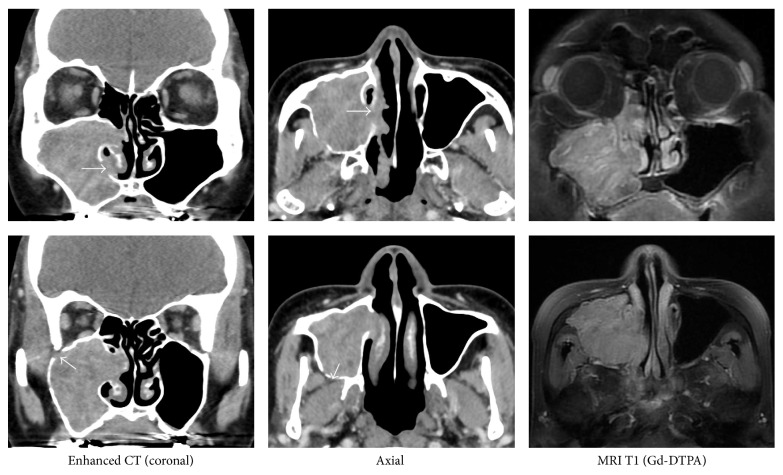
Figure 2.
CT and MRI Imaging. The contrast CT shows bone defects in the anterior and medial walls of the maxillary sinus. There are no indications that the thickening of the bone should be suspected as the site of origin. MRI T1 (Gd-DTPA) shows secondary maxillary sinusitis and a serpentine cerebriform filamentous structure, but there is no mass to otherwise indicate a possible origin.

