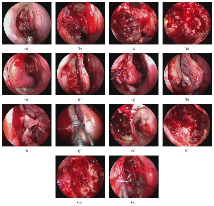
Figure 3.
Endoscopic Modified Medal Maxillectomy (EMMM). (a) The uncinate process was resected, and an IP deviating from the maxillary sinus was observed. (b) The uncinate process and ethmoidal bulla were resected, and the maxillary sinus was opened. A tumor was found to be filling the maxillary sinus. (c) Inverted papilloma in the maxillary sinus was debulked. All of the IPs were adhering to the posterior wall. (d) The anterior and inferior walls of the maxillary sinus were not visible when viewed from the fontanelle of the maxillary sinus with a 70-degree endoscope. (e) From the anterior portion of the inferior turbinate behind the pyriform aperture, the mucosa was incised towards the floor of nasal cavity (black line). (f) The mucosa of inferior turbinate was detached. The mucosa on the inferior meatus was detached to expose the conchal crest. (g) The conchal crest was cut with a chisel, the inferior turbinate bone was disengaged, and the inferior turbinate was moved medially. (h) When the inferior turbinate was moved medially, the conchal crest and the inferior end of the nasolacrimal duct (∗) could be observed. (i) The nasolacrimal duct was identified, and the IP that was deviating to the inferior meatus was resected. (j) The lacrimal process of the inferior turbinate, frontal process of the maxilla, and inferior portion of the lacrimal bone were drilled with 2.5 mm Curved Diamond DCR Bur (Medtronic), in order to make the superior portion of the lacrimal duct visible. (k) The inside of the maxillary sinus was visible from the inferior meatus side. (l) Inverted papilloma was observed on the anterior and inferior walls. (m) All of the IPs found on the anterior and inferior walls were resected with the 70-degree endoscope. (n) The pyriform aperture and mucosa of the inferior turbinate were sutured and the surgery was completed.

