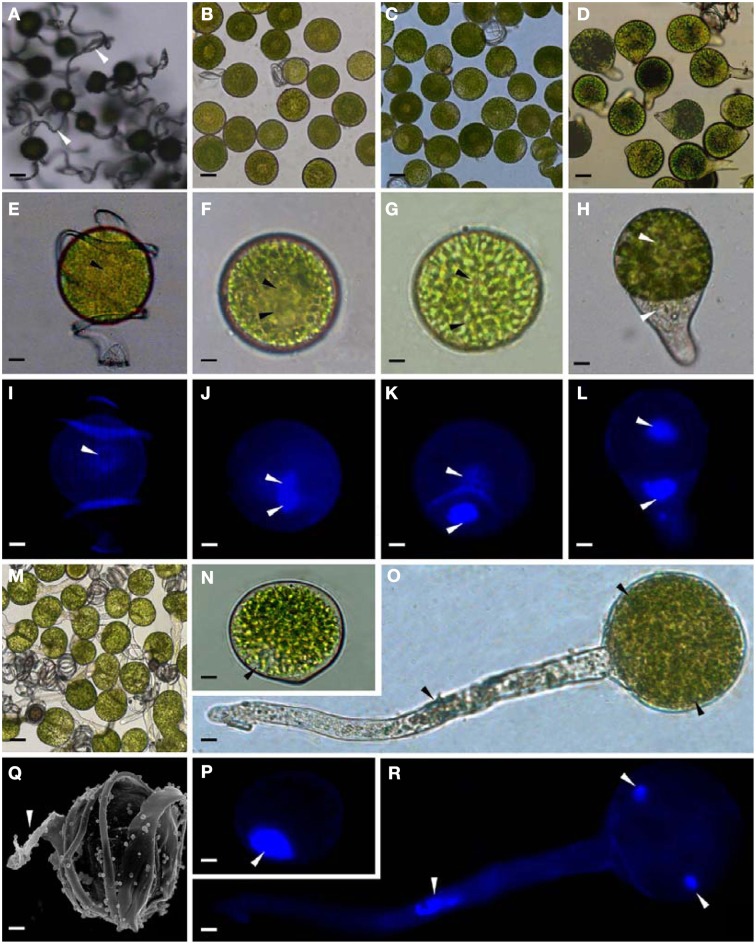
Figure 2.
Morphology of mature and germinating spores from E. arvense. (A,E,I,N,P,Q) Mature spores (MS). (A) MS groups, arrows show elaters, bar = 20 μm; (E,I) nucleus center-localized MS with elaters under bright-field microscope (BFM) and fluorescence microscope (FM), arrows show nuclei, bar = 5 μm; (N,P) nucleus-migrated MS without elaters under BFM and FM, arrows show nuclei, bar = 5 μm; (Q) MS with elaters under scanning electron microscope, arrows show elaters, bar = 8 μm. (B,F,J) Rehydrated spores (RS). (B) RS groups, bar = 15 μm; (F,J) RS under BFM and FM, arrows show nuclei, bar = 5 μm. (C,G,K) Double-celled spores (DCS). (C) DCS groups, bar = 15 μm; (G,K) DCS under BFM and FM, arrows show nuclei, bar = 5 μm. (D,H,L) Germinated spores (GS). (D) GS groups, bar = 15 μm; (H,L) GS under BFM and FM, arrows show nuclei, bar = 5 μm. (M,O,R) Spores with protonemal cells (SPC). (M) SPC groups, bar = 15 μm; (O,R) SPC under BFM and FM, arrows show nuclei, bar = 5 μm.

