Fig. 3.

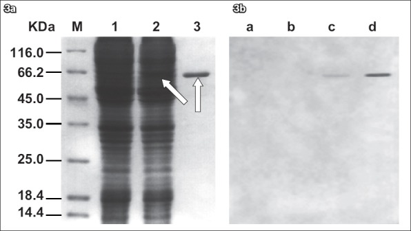
(a) SDS-PAGE of the lysates of the non-infected Sf9 cells and the baculovirus-infected Sf9 cells. Lane 1: lysates of the non-infected Sf9 cells; lane 2: lysates of the baculovirus-infected Sf9 cells; lane 3: purified recombinant gG321–580His. The lysates were separated using 12% SDS-PAGE. Additional bands (arrows) were observed in lanes 2 & 3 after Coomassie blue staining. A protein marker is shown at the left part of the Coomassie-stained gel (lane M). (b) Western blot analysis of the recombinant gG321–580His. Lane a: protein marker; lane b: non-infected Sf9 cells culture; lane c: baculovirus-infected Sf9 cells culture; lane d: purified recombinant gG321–580His. A single band was observed in both lanes c & d, which showed that the recombinant protein gG321–580His was able to react with the specific mouse anti-gG-2 monoclonal antibody. No Western blot signal was observed in lanes a & b (negative controls), indicating that the recombinant protein gG321–580His has similar functions with its natural counterpart.
