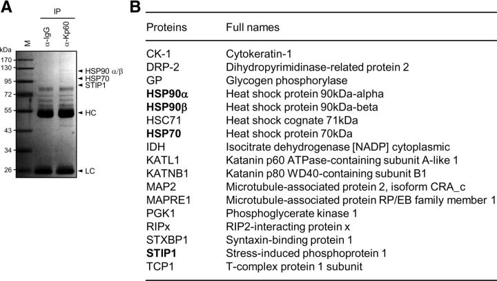
Figure 3.
Identification of katanin-p60-binding proteins. A, Lysates from cortical neurons at 7 DIV were subjected to immunoprecipitation with IgG or anti-katanin-p60 antibody. Precipitated proteins were subjected to SDS-PAGE followed by staining with Coomassie Brilliant Blue R250. They were also subjected to mass spectrometry for the identification of katanin-p60-binding proteins. The arrowheads indicate some of the proteins identified by the mass analysis. HC and LC indicate IgG heavy and light chains, respectively. M denotes size markers. B, List of proteins identified as katanin-p60-interacting proteins. Proteins bound to anti-katanin-p60 antibody were precipitated and analyzed by LC-MS/MS.

