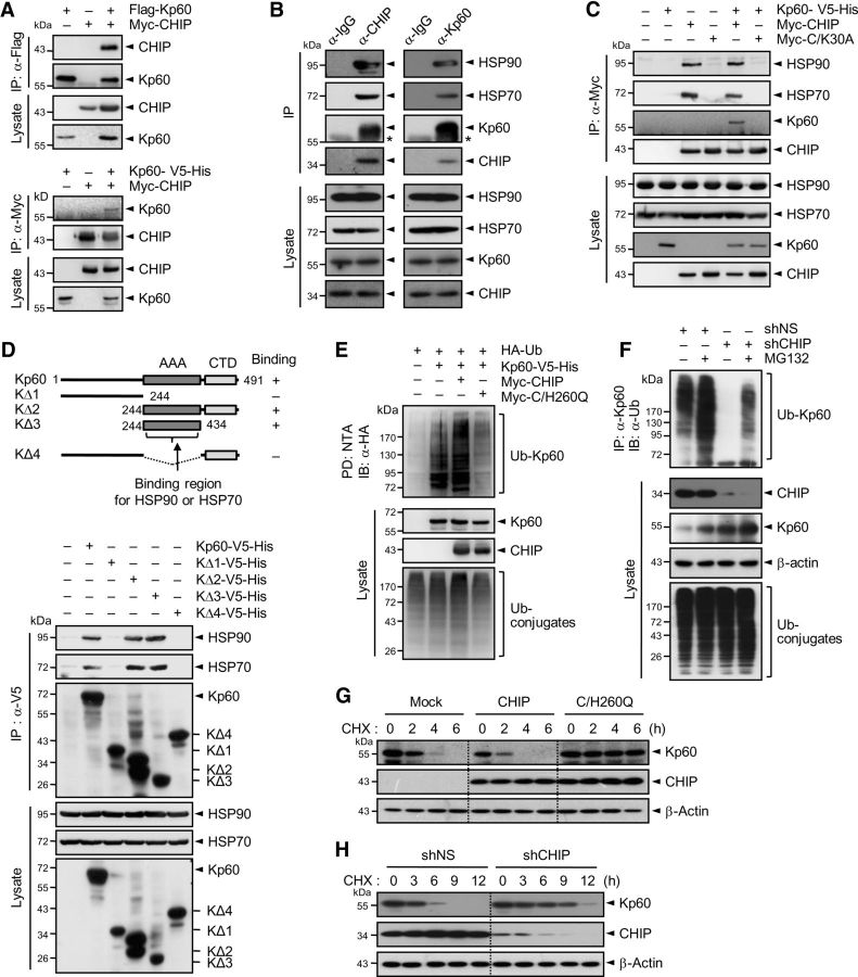
Figure 4.
CHIP interacts with, ubiquitinates, and destabilizes katanin-p60. A, Flag-Kp60 (top) or Kp60–V5-His (bottom) were expressed in HEK293T cells with Myc-CHIP. Cell lysates were subjected to immunoprecipitation with anti-Flag or anti-Myc antibody followed by immunoblot with anti-Myc or anti-Flag antibody, respectively. B, Lysates of cortical neurons at 7 DIV were immunoprecipitated with anti-CHIP (left) or anti-Kp60 antibody (right) followed by immunoblot with respective antibodies. C, Kp60–V5-His was expressed in HEK293T cells with Myc-tagged CHIP or C/K30A. Cell lysates were immunoprecipitated with anti-Myc antibody. D, Deletions of katanin-p60 (KΔ1–KΔ4), prepared as in Figure 2B, were expressed in HEK293T cells (top). Cell lysates were subjected to immunoprecipitation with anti-V5 antibody followed by immunoblot with anti-HSP90, anti-HSP70, or anti-V5 antibody (bottom). In the top panel, whether the proteins bind to each of the deletions or not was shown as “+” or “−,” and the binding region was indicated by the arrow. E, HA-Ub and Kp60–V5-His were expressed in HEK293T cells with Myc-tagged CHIP or C/H260Q. Cell lysates were pulled down with NTA resins. F, Neuro2A cells were transfected with shNS or shCHIP. After culturing for 48 h, cell lysates were immunoprecipitated with anti-Kp60 antibody. G, Kp60–V5-His was expressed in Neuro2A cells with Myc-tagged CHIP or C/H260Q. After culturing for 24 h, cells were treated with cycloheximide. H, Neuro2A cells were transfected with shNS or shCHIP. Cells were treated as in G. Note that in A, E, and F cells were incubated with 10 μm MG132 for 4 h before the preparation of cell lysates.

