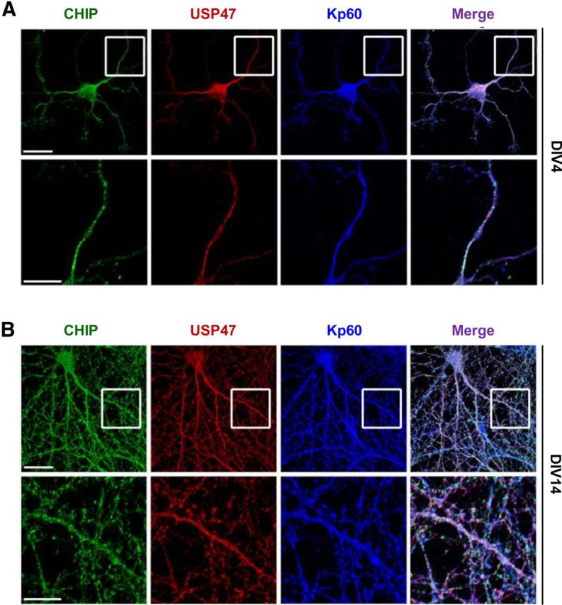
Figure 6.

USP47 colocalizes with katanin-p60 and CHIP. A, B, Hippocampal neurons were cultured for 4 DIV (A) and 14 DIV (B) on slide glasses. Cells were then fixed; stained with anti-CHIP (green), anti-USP47 (red), and anti-katanin-p60 antibodies (blue); and observed under a fluorescent microscope. The bottom panels show the magnified views of the boxed regions of the top panels. Scale bars: top panels, 20 μm; bottom panels, 10 μm.
