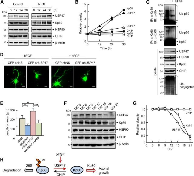
Figure 8.
bFGF increases the expression of USP47 and katanin-p60. A, Cortical neurons at 3 DIV were treated with 60 ng/ml bFGF followed by immunoblot analysis. B, The band intensities seen in A were determined by using a densitometer. Similar data were obtained in three independent experiments. The open and closed symbols show without and with bFGF, respectively. C, Cortical neurons were incubated for 36 h with and without bFGF (60 ng/ml). Cell lysates were immunoprecipitated with anti-katanin-p60 antibody followed by immunoblot with anti-Ub or anti-Kp60 antibody. D, GFP-shNS and GFP-shUSP47 were expressed in hippocampal neurons at 3 DIV. After culturing them with and without bFGF (60 ng/ml) for 48 h, cells were fixed and observed. Statistical analysis was performed by using Student's t test. **p < 0.005; ***p < 0.001. Scale bars, 20 μm. E, The lengths of axons were determined as in Figure 7B. Error bars indicate SD. F, Cortical neurons were cultured and immunoblotted. G, The band intensities seen in F were determined by using a densitometer. Similar data were obtained in three independent experiments. H, A model for the control of axonal growth by USP47 and CHIP.

