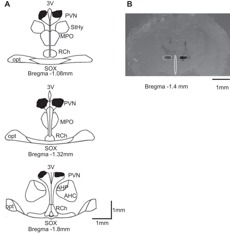
Fig. 4.
Schematic representation of coronal sections throughout the rat hypothalamus. A: shaded areas indicate brain regions exposed to dye used to mark the injection sites bilaterally. The shape of each area was determined by overlaying tracings of the outermost diffusion area of injected dye (50 nl) observed on each section through the PVN. B: representative single bilateral injection of dye within the PVN (50 nl of 2% Chicago blue dye each). 3V, third cerebral ventricle; StHy, striohypothalamic nucleus; MPO, medial preoptic nucleus; RCh, retrochiasmatic area; opt, optic tract; SOX, supraopticdecussation; AHP/AHC, anterior hypothalamic area.

