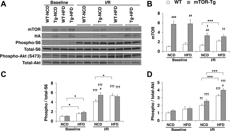
Fig. 4.
Overexpression of cardiac mTOR induces functional activation of both mTORC1 and mTORC2 in post-I/R hearts. A: representative immunoblots of mTOR signaling molecules in hearts subjected to the ex vivo Langendorff perfusion model. Baseline hearts were harvested after 15 min of equilibration perfusion ex vivo. I/R hearts were harvested after a course of baseline conditions followed by 20-min ischemia and then 40-min reperfusion. Immunoblot analysis was performed with the indicated antibodies. Blots are representative of six independent experiments. Densitometric quantitative analyses of mTOR (B), phospho-S6 (C), and phospho-Akt (D) were normalized to baseline levels of WT-NCD hearts in each experiment. n = 6 baseline hearts and 12 I/R hearts. *P < 0.05 and ***P < 0.001, NCD vs. HFD; #P < 0.05, ##P < 0.01, and ###P < 0.001, WT vs. mTOR-Tg hearts; †P < 0.05, ††P < 0.01, and †††P < 0.001, baseline vs. I/R (by Student's t-test).

