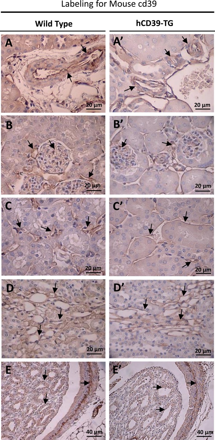
Fig. 1.
Immunoperoxidase labeling for mouse cd39 protein in the kidneys of wild-type (WT, left) and transgenic (hCD39-TG, right) mice. Labeling for mouse cd39 was performed on paraffin-embedded kidney sections. Magnification is indicated in each panel. A and A′: blood vessels; B and B′: glomeruli and glomerular blood vessels; C and C′: perivascular blood vessels and capillaries; D and D′: vascular bundles at the corticomedullary region; E and E′: IMCD cells in papilla and on pelvic wall.

