Fig. 3.
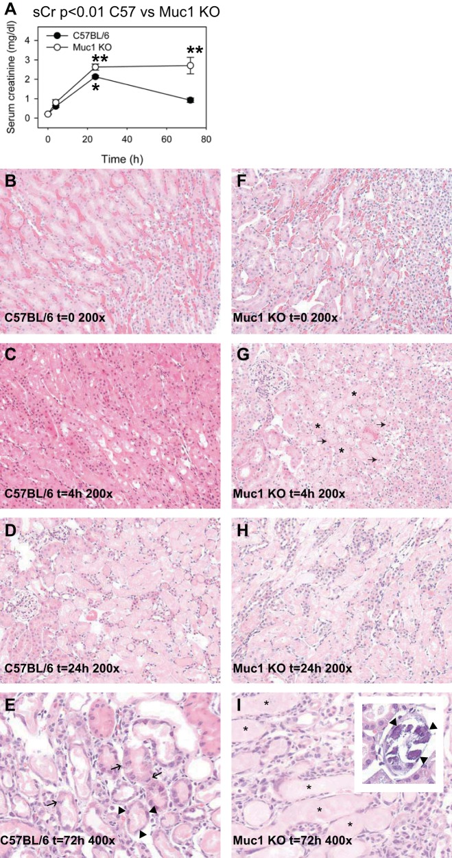
Muc1 protects kidney function and morphology during kidney IRI. Kidneys of Muc1 KO mice and congenic control C57BL/6 mice were subjected to IRI for 19 min and recovery for 0–72 h. A: blood was collected at the time of death and assayed for serum creatinine (sCr). Profiles for sCr between Muc1 KO mice (open circles) and congenic wild-type mice (closed circles) were significantly different by two-way ANOVA (n = 3–6 mice at each time point, P < 0.01). Values at 72 h (means ± SE) were different between Muc1 KO and wild-type mice (P < 0.001). Values at 24 h in control mouse kidneys were significantly higher than at t = 0 (*P < 0.01). Values for Muc1 KO mouse kidneys at both 24 and 72 h were significantly higher than at t = 0 (**P < 0.001). B–I: one kidney from each mouse was fixed and stained with hematoxylin and eosin and scored for tubule damage as described in the text. B–H: representative images (×200) are shown for control (B–D) and Muc1 KO (F–H) kidneys at 0, 4, and 24 h of recovery, respectively. G: karyorrhexis and karyopyknosis (arrows) and luminal casts (*) were especially evident in Muc1 KO kidneys at t = 4 h. E and I: images (×400) from 72 h of recovery are shown for control C57BL/6 (E) and Muc1 KO (I) mice. E: mitotic figures (arrows) and flattened cells in recovering tubules (arrow heads) were observed in control mouse kidneys at 72 h. I and inset: luminal casts (*) and calcium phosphate precipitates (arrowheads) were observed in Muc1 KO kidneys at 72 h.

