Fig. 2.
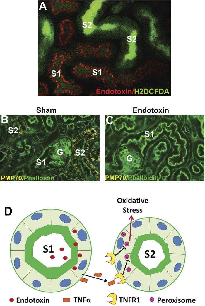
Tubular cross talk in sepsis-induced acute kidney injury (AKI). In A, intravital 2-photon microscopy reveals that systemically administered fluorescent endotoxin (red) accumulates in S1 tubules via Toll-like receptor 4 (TLR4)-mediated endocytosis. Neighboring S2 segments (but not S1) exhibit severe oxidative stress (H2DCFDA, green). In B, fluorescence microscopy of fixed kidneys shows abundant peroxisomes (PMP70, yellow) in S2 segments (but not S1) under sham conditions. G denotes glomeruli, and green is FITC-phalloidin staining of actin. In C, S2 segments show reduced peroxisomal staining 4 h after endotoxin injury. The cartoon in D depicts S1 as a sensor of danger molecules such as endotoxin in the filtrate. S1 then signals S2 through cytokines such as TNF-α which act on cognate receptors such as TNF-α receptor 1 (TNFR1) present only on S2. In cases of severe stress, this signaling causes early peroxisomal damage and oxidative stress.

