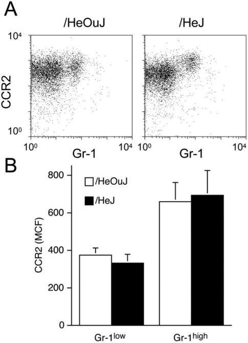
Figure 3.
CCR2 staining on blood monocytes from /HeOuJ and /HeJ mice. A. Blood leukocytes were isolated from /HeOuJ and /HeJ mice 1 day after sponge insertion. The figure shows Gr-1 and CCR2 staining on gated blood monocytes (SSClow F4/80+). B. Median channel fluorescence of CCR2 stained blood monocytes, according to Gr-1 expression. Graphs show the values of the means and SD n > 5 animals per group.

