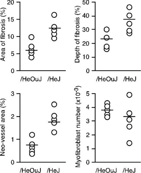
Figure 6.
Increased fibrosis and neovascularization in wounds in TLR4 deficient mice. Histomorphometric analysis of sponges removed 21 days after insertion in /HeOuJ or /HeJ mice. Graphs show area of fibrosis, maximum depth of fibrosis, area of the wound invaded by neovessels, and the number of myofibroblasts in the wound for each animal. Open circles show values from individual animals, and the group means are represented with a horizontal bar. p < 0.05 between strains, except for the number of myofibroblasts (P > 0.05), Mann–Whitney's U-test, n = 5 mice per group.

