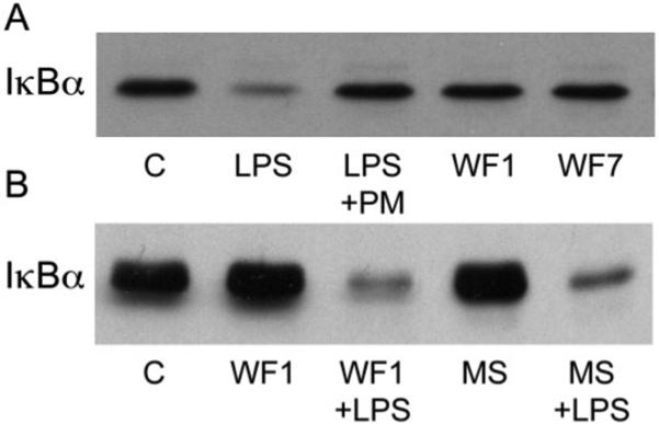
Figure 7.
Wound fluids do not induce IκBα degradation or interfere with NFκB activation by lipopolysaccharide (LPS). A. Murine J774.A macrophages were untreated (Control: C) or exposed to LPS (100 ng/mL) ± polymyxin B (PM, 50 ng/mL), or day 1 (WF1) or day 7 (WF7) wound fluids (50%, v : v) for 30 minutes in culture medium containing 1% FBS. Cell lysates were immunoblotted for IκBα. B. Cells were cultured with day 1 wound fluids (WF1) or normal mouse serum (MS) at 50% (v : v), with or without LPS (100 ng/mL), as described under (A) and assayed for IκBα content. Intervening noncontributory lanes were removed from the image for clarity.

