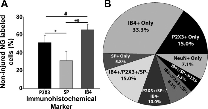
Fig. 1.
Immunohistochemical representation of P2X3, isolectin B4 (IB4), and substance P (SP) in nodose ganglion (NG) neurons. A: following staining of the immunohistochemical markers P2X3, SP, and IB4, the bar graph demonstrates that all three were well represented in the NG, with the majority being IB4+ (IB4 vs. P2X3, #P < 0.05; IB4 vs. SP, **P < 0.001; P2X3 vs. SP, *P < 0.01). Values are means ± SD; n = 3 rats and 6 ganglia (one-way ANOVA with Tukey post hoc t-tests). B: pie graph depicting all possible combinations of the molecular targets in the NG. Note that neurons that were IB4+ only were the most prevalent, and most NG neurons were labeled with at least one of the three cellular targets examined.

