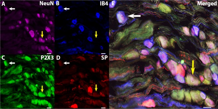
Fig. 2.
Quadruple immunohistochemical staining in the NG. A: NeuN. B: IB4. C: P2X3. D: SP. E: merged. A confocal image displays the typical staining within the NG. NeuN was used to label all neurons. Different histochemical combinations include neurons that were IB4+, P2X3+, but SP− (white arrows), and neurons that were IB4−, P2X3+, and SP− (yellow arrows). Scale bar indicates 25 μm (×20 objective).

