
Fig. 3.
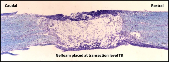
Spinal transection injury at T8. An 18-μm-thick sagittal section stained with the Kluver-Barrera method illustrates a complete spinal transection at T8. Gelfoam is placed in the lesion cavity to prevent contact from between rostral and caudal spinal cord tissue. The scale bar indicates 500 μm (×4 objective).