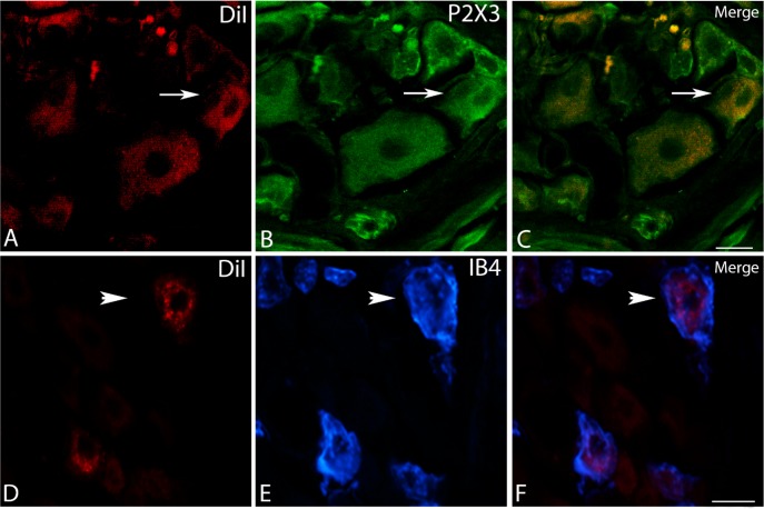
Fig. 8.
P2X3-ir and IB4 binding in bladder-traced NG neurons after transection. A confocal image illustrates a DiI+ neuron in A that is also immunoreactive for P2X3 in B (white arrows). C: demonstration of the overlay. An image from the inverted Nikon microscope illustrates a DiI+ neuron in D that also binds IB4 in E (white arrowhead). F: demonstration of the overlay. One example of each is displayed. In both images, the scale bar indicates 25 μm (×20 objective).

