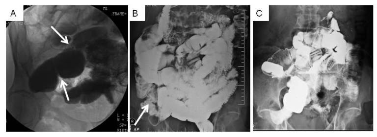
Figure 1.
Small bowel radiographs of Patient 1 documenting SBO resolution over time. Arrows note areas of obstruction. (A) Before therapy in 2011: incomplete SBO due to adhesions visualized by X-ray showing dilation of the proximal mid ileum. (B) Twelve months after therapy in 2012: mild stricture at the terminal ileum with no other small bowel abnormalities. (C) After 40 h of therapy: normal small bowel series X-ray in 2012.

