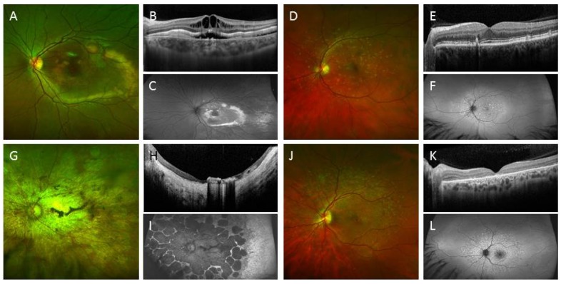
Figure 6.
Clinical images of various types of inherited retinal diseases. (A) Colour photograph of the macula of a 10-year-old boy showing multifocal vitelliform lesions resulting from homozygous deletion of exon 2–6 of the BEST1 gene; (B) Optical coherence tomography (OCT) shows intraretinal cystic change with sub-retinal fluid and vitelliform deposits; (C) Increased fundus autofluorescence was noted in the area of vitelliform deposits; (D) Colour photograph of the macula of a 57-year-old male showing yellow deposits due to pattern dystrophy of the retinal pigment epithelium (RPE); (E) OCT shows deposits above and below the RPE; (F) Multifocal hyper autofluorescent lesions are seen; (G) Colour photograph of the macula of a 56-year-old female showing extensive macular atrophy with cone-rod dystrophy due to two missense mutations in the ABCA4 gene (c.2915 C > A and c.3041 T > G); (H) OCT shows severe retinal and choroidal atrophy with pigment migration into the fovea; (I) Extensive RPE loss resulting in wide-spread hypo autofluorescent lesions; (J) Colour photograph of the macula of a 32-year-old male showing retinal flecks with mild cone dysfunction due to two pathogenic mutations in the ABCA4 gene (c.4139 C > T and c.6079 C > T); (K) OCT shows outer retinal layer loss to retinal atrophy; (L) Retinal flecks were hyper autofluorescent.

