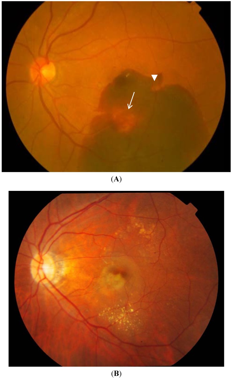
Figure 1.
Fundus photographs showing the two clinical patterns of polypoidal choroidal vasculopathy: Hemorrhagic (A) and exudative (B). An orange red nodule suggestive of a polyp (white arrow) is seen in an extrafoveal position, while a notch in the hemorrhagic pigment epithelial detachment (white arrowhead) suggests the presence of another polyp.

