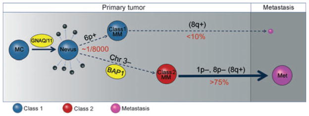
Figure 3.

Summary of major molecular events in uveal melanoma progression. The earliest known and perhaps initiating event is an activating mutation in GNAQ or GNA11, presumably in a normal uveal melanocyte (MC), which may function primarily to trigger inappropriate cell cycle re-entry through activation of the MAPK and perhaps other pathways. Usually the mutant cell clone does not progress to melanoma, but rather undergoes senescence resulting in a nevus, or is eliminated by apoptosis or immune surveillance (small black spheres). Less than one in 8000 nevi progress beyond this stage (Singh et al., 2005b). The rare tumor that progresses does so along one of the two pathways characterized by distinct gene expression profiles (GEP). The GEPs of normal uveal melanocytes and nevi are very similar to that of class 1 uveal melanomas (blue spheres) (Chang et al., 2008), which have a low risk of metastasis (small purple sphere). Melanomas that acquire the class 2 GEP (class 2 MM) have a very high risk of metastasis (large purple sphere). Class 1 tumors often exhibit 6p gain and 8q gain, but have less overall aneuploidy than class 2 tumors, which often exhibit 1p loss, 8p loss, and 8q gain. 8q gain is more common in class 2 tumors, but is also seen in class 1 tumors, so this may be a late event (Parrella et al., 1999). The class 2 GEP is strongly associated with mutation of BAP1, located at 3p21, and loss of the other copy of chromosome 3, suggesting that bi-allelic loss of BAP1 is a key step in uveal metastasis (Harbour et al., 2010). Metastatic tumors (purple spheres) have a distinct gene expression profile that is more similar to that of class 2 than that of class 1 primary tumors (author’s unpublished data). MC, melanocyte; MM, malignant melanoma; Met, metastasis.
