Figure 1.
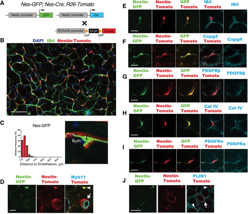
Nestin-Cre and Nestin-GFP identify perivascular cells in WAT. (A) Schematic of the genetic tools in Nes-GFP; Nes-Cre; R26-Tomato dual-reporter mice used in this figure. GFP and Cre are expressed from distinct nestin-driven transgenes. Cre acts on a Cre/lox-dependent R26 knock-in fluorescent Tomato reporter, which serves as a lineage trace. (B) Epifluorescence of Nes-Cre/Tomato lineage tracing in visceral WAT, imaged by whole-mount with isolectin-IB4 staining for capillary endothelial cells. (C) Measurement of the distance between DAPI+ nuclei of individual Nes-GFP+ cells (n = 167) and the nearest IB4+ capillary membrane, as shown in the example at the right. A distance <10 µm means the cell is on the abluminal surface of the capillary. (D) Epifluorescence of Nes-GFP and Nes-Cre/Tomato plus immunofluorescence staining of Myh11 in vascular smooth muscle cells. The GFP/Tomato reporters are coexpressed in pericyte-like cells (arrowhead). Tomato also identifies adventitial cells in which Nes-GFP is not expressed (arrow). (E–I) Epifluorescence of Nes-GFP and Nes-Cre/Tomato plus IB4 labeling for endothelial cells (E), immunofluorescence staining of Cspg4 or PDGFRβ for pericytes (F,G), immunofluorescence staining of collagen IV for basement membrane (H), and immunofluorescence staining of PDGFRα (I). (J) Epifluorescence of Nes-GFP and Nes-Cre/Tomato plus immunofluorescence staining of Perilipin (PLIN1) for adipocytes. Tomato identifies rare adipocytes (arrow) that do not express Nes-GFP. A Tomato+GFP+ pericyte-like cell is also shown (arrowhead). Bars: B, 100 µm; D, 30 µm; E–I, 20 µm; J, 30 µm.

