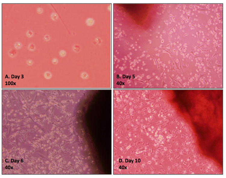
Figure 1. Phase contrast micrographs of cell isolates from a mouse calvarium.

A) Day 3 image demonstrating the migration of cuboidal-shaped cells from harvested bone. B) Day 5 image demonstrating adherence of the cells to the plastic culture plates and acquisition of a stromal cell morphology. C) Day 7 and D) Day 10-12 images demonstrating 50% and 80-100% confluency, respectively. Cells were subsequently passaged into 25mm2 flasks.
