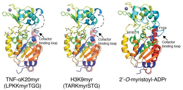
Figure 5. Crystal structure of SIRT2 in complex with TNFα-K20myr, H3K9myr or 2′-O-myristoyl-ADP-ribose.
Overall structural features of SIRT2 in the presence of myristoylated substrates or product. The cofactor binding loop was not visible in the SIRT2 – TNFα-K20myr or SIRT2 – H3K9myr structures and is shown as a dashed line for clarity. Also highlighted in the 2′-O-myristoyl-ADPr structure are the locations and orientations of Tyr104 and the catalytic histidine, His187.

