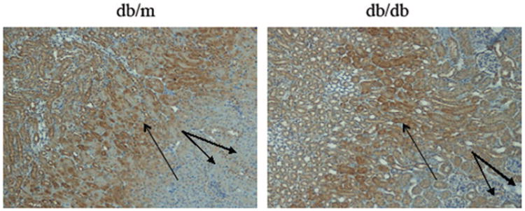
Figure 2.

Pimonidazole immunohistochemical staining of the kidney of db/m and db/db mouse. Strong pimonidazole staining of renal tubules was observed mainly in the OM (single arrows) both from db/m and db/db mouse. Weaker staining was seen in the cortex of both db/db and db/m mice (double arrows, 100×).
