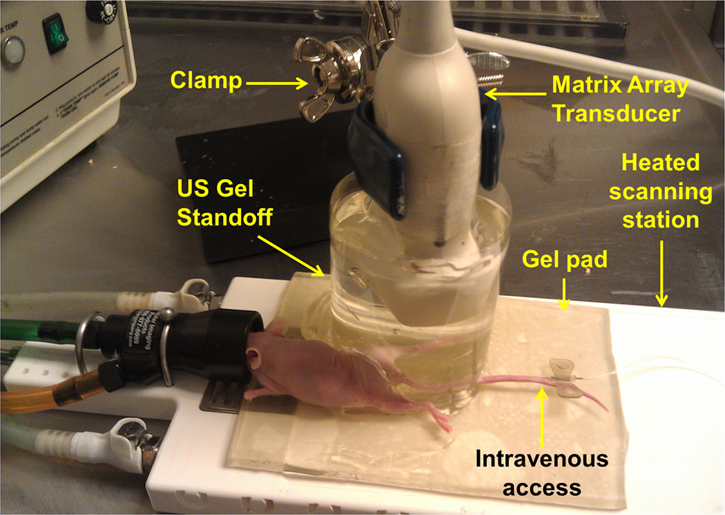
Figure 1.
Photograph of the imaging setting for three-dimensional (3D) ultrasound molecular imaging (USMI) using a clinical matrix array transducer in mice. To bring the subcutaneous human colon cancer xenograft implanted on the hind limb beyond the near field zone of the transducer, the transducer was embedded in a custom standoff, which was comprised of a column of pre-warmed ultrasound gel contained within a plastic cylindrical chamber. All mice were kept under inhalation anesthesia during scanning and body temperature was kept constant by placing them on a gel pad on a heated scanning station. Note, a needle was placed in one of the two tail veins to allow intravenous administration of contrast microbubbles.

