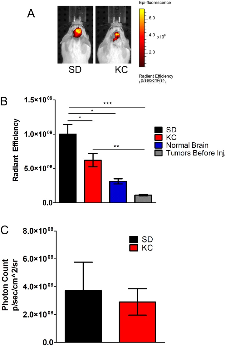
Fig 2. In vivo imaging of hypoxia.
(A) The fluorescent probe HypoxiSense 680 was used to analyze hypoxia in vivo 21 days following tumor implantation. (B) Fluorescent signal was quantitated from tumor bearing mice (N = 5; *p < 0.05). Animals were imaged prior to injection to analyze tissue autofluorescence (“Before injection”; N = 5; ***p < 0.001). Non-tumor bearing mice were injected to analyze non-specific binding (“Normal Brain”; N = 2; *p < 0.05). (C) Tumor bioluminescence imaging showed no significant difference between SD and KC (N = 5).

