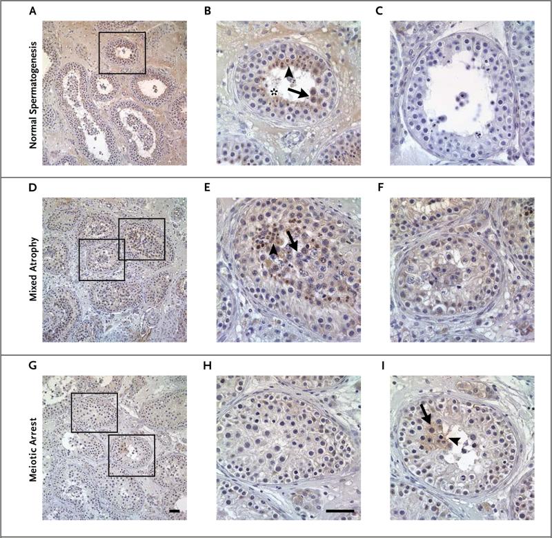
Figure 3. Immunohistochemical Detection of TEX11 in Testicular Tissue Sections Obtained from a Man with Normal Spermatogenesis, a Man with Mixed Testicular Atrophy, and a Man with Meiotic Arrest.
In a man with normal spermatogenesis (Panels A and B), TEX11 was highly expressed in late spermatocytes (arrow), round spermatids (arrowhead), and elongated spermatids (asterisk). Testicular tissue stained with IgG antibody was used as a negative control (Panel C). TEX11 was also present in tubules with remaining spermatogenesis in a patient with mixed testicular atrophy. TEX11 expression was observed in late spermatocytes (arrow) and round spermatids (arrowhead) (Patient 6) (Panels D and E). Seminiferous tubules containing only less-differentiated germ cells and Sertoli cells show no TEX11 expression (Panel F). In a man with complete meiotic arrest (Patient 4), germ cells did not have TEX11 expression in the majority of tubules (Panels F, G, and H). However, TEX11 staining was observed in rare seminiferous tubules with remaining late spermatocytes (arrow) and round spermatids (arrowhead) (Panel I). Scale bars indicate 50 μm.

