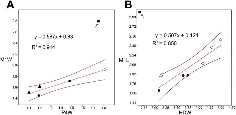
Fig 3. Simple linear regression plots (OLS) with 95% confidence limits (blue lines) of dental and postcranial measurements of mystacinids (Table 1).
A, Posterior upper premolar width (P4W) against first upper molar width (M1W). B, Distal humerus width (HDW) against first upper molar length (M1L). Square in each graph indicates M1 of Mystacina miocenalis plotted against value for the largest specimens of P4 and HD (respectively) for mystacinids previously recovered from St Bathans [7]. Mystacina tuberculata (filled circle), M. robusta (open circle), Icarops paradox (filled triangle), I. aenae (open triangle).

