
Fig 10. RGC loss in GPx4+/− and GPx4+/+ mice after 7 days of NMDA or vehicle treatment.
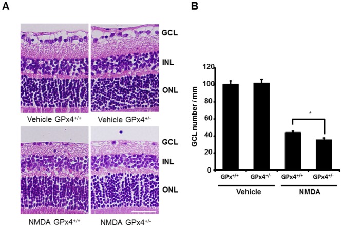
(A) Hematoxylin and eosin staining of retinal sections. Scale bar, 30 μm. (B) The number of cells in GCL were compared between of GPx4+/− and GPx4+/+ mice. Data are mean ± SEM (n = 9–10). *p < 0.05.