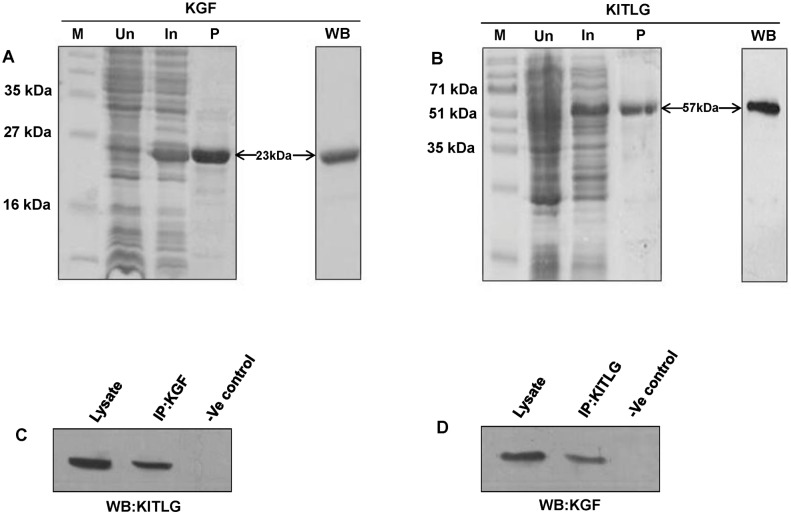
Fig 2. SDS-PAGE and validation of buffalo KGF and KITLG proteins.
SDS-PAGE profiles of buffalo KGF (A) and KITLG (B) proteins showing their resolved chromatographic fractions. M, Un, In and P denote marker, uninduced, induced and purified protein samples. The purified KGF and KITLG proteins were validated using western blot (WB) with anti-His (panel A) and anti-GST antibodies (panel B), respectively. The 23 and 57 kDa bands correspond to the purified KGF (His-tagged) and KITLG (GST tagged) proteins. (C and D) Co-immunoprecipitation of KGF and KITLG proteins. (C) The buffalo KGF and KITLG protein interactions were confirmed by immunoprecipitation of the tissue (ovary) lysate with anti-KGF antibody followed by immunoblotting with anti-KITLG IgG and detected a specific band of 31 kDa corresponding to KITLG protein. (D) In the reciprocal assay, a band of 22 kDa was detected on immunoprecipitation with anti-KITLG antibody followed by western blotting with anti-KGF IgG. The tissue lysate and resin in both C and D panels denote the positive and negative controls, respectively.

