
Figure 5.
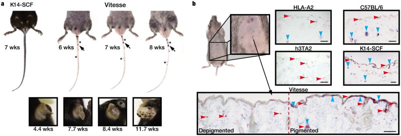
SCF overexpression is associated with formation of pigmented lesions. (a) Rapid depigmentation is accompanied by the gradual development of repigmenting lesions. Pigmented lesions increased in pigmentation (arrow) and number (asterisks) over time (b) Epidermal repigmentation underneath the pelage of Vitesse mice. Expression of melanocyte marker, Trp-1 (blue) and pan-T cell marker, CD3 (red) were observed in the skin of C57BL/6, HLA-A2, and K14-SCF mice. Melanocytes were absent from skin of h3TA2 and Vitesse mice except within pigmented lesions, where a near original melanocyte distribution was accompanied by infiltrating T cells. n=3. Scale bar = 0.2mm.