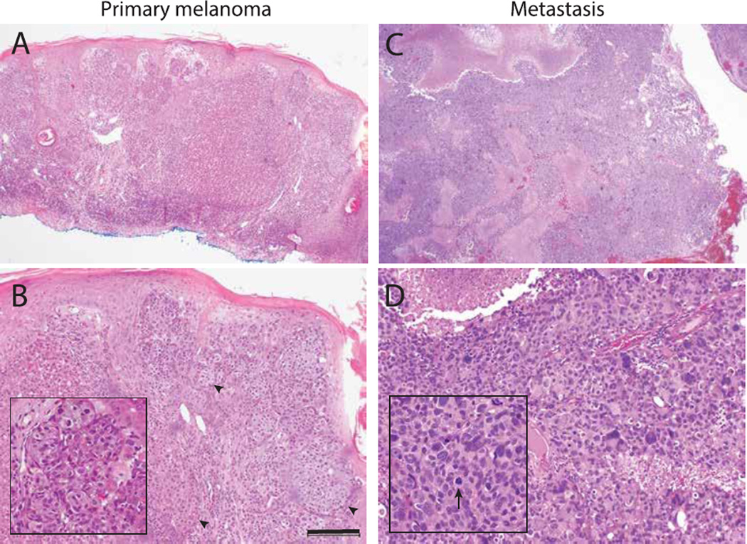
Figure 1.
Histology of primary and metastatic melanoma. (A) The primary cutaneous lesion is an asymmetric cellular tumor filling the dermis with lack of maturation at the base of the lesion. (B) The primary lesion also shows epithelial hyperplasia and some nests comprised of large and epithelioid cells with abundant cytoplasm (arrowheads). Inset highlights a nest of epitheliod melanocytes. (C) The metastatic cerebellar lesion shows a densely cellular neoplasm with zones of necrosis and hemorrhage. (D) There is marked pleomorphism at higher magnification. Arrows indicates an aberrant mitotic figure. Scale bar=200µm.

