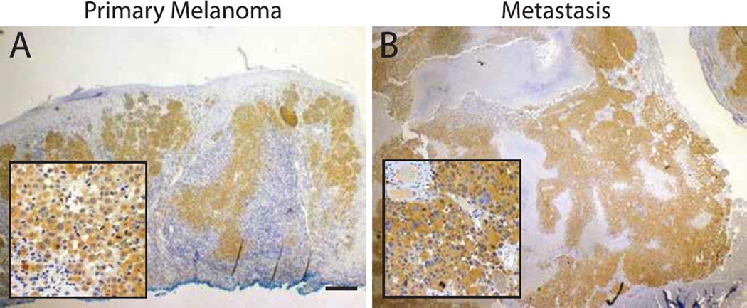
Figure 2.
BRAF immunohistochemistry. (A) Melanoma biopsy shows diffuse cytoplasmic staining by the BRAF-V600E specific antibody (VE1) supporting the presence of underlying BRAF-V600E mutation. Inset shows individual B-Raf-mutant melanocytes invading the dermis. (B) Cerebellar lesion shows strong BRAF-V600E reactivity by the infiltrating tumor cells. Scale bar=200µm.

