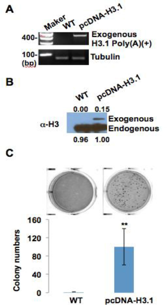
Figure 2. Polyadenylation of H3.1 facilitates colony formation of BEAS2B cells on soft agar.
(A) RT-PCR results. Canonical histone H3.1 cDNA was inserted into pcDNA vector, which contains poly(A) signal and thus generates polyadenylated H3.1 mRNA. pcDNA-H3.1 vector was stably transfected into BEAS2B cells. Total RNA was extracted from each cell line and then converted to cDNA using oligo dT primers. Exogenous polyadenylated H3.1 mRNA was then amplified by PCR with specific primers for H3.1 and vector. (B) Western blot shows exogenous H3.1 protein generated from ectopically expressed polyadenylated H3.1 mRNA. (C) BEAS2B cells (WT) and pcDNA-H3.1-expressing cells were plated in 0.35% soft agar, and cultured for 4 weeks. The data shown are the means ± S.D. from experiments performed in triplicate. ** p<0.01.

