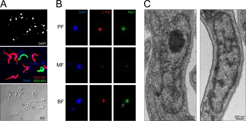
Fig. 2.
Coat formation by a single mVSG in metacyclics is followed by dispersal of the nucleolus. (A) Metacyclic cells were purified from culture also containing procyclics and epimastigotes on 0.1 mm diameter zirconia/silica beads column prepared in a Pasteur pipette in BBSG buffer (50 mM bicine, 50 mM NaCl, 5 mM KCl, 70 mM glucose). IF assay was performed with paraformaldehyde-fixed cells with the anti-VSG 397 rabbit serum (diluted 1:5,000) and anti-VSG 653 rat serum (diluted 1:500) and Alexa Fluor 488-conjugated goat anti-rabbit and Alexa Fluor 594-conjugated goat anti-rat secondary antibodies (Invitrogen), diluted 1:1,000. Note that some metacyclics are not recognized by either anti-VSG antibodies. (B) IF assay with affinity-purified anti-Pol I antibodies and L1C6 mouse monoclonal antibody was performed on PF, purified MF, and T.brucei Lister 427 single marker BF cells as described previously [3]. Arrow points to the ESB in BF. (C) Transmission EM of nucleus of a cell without a VSG coat (left) and a VSG-coated cell (right). The nucleolus is easily identifiable in the nonmetacyclic nucleus as an electron-dense sphere and it is not discernible in the metacyclic nucleus.

