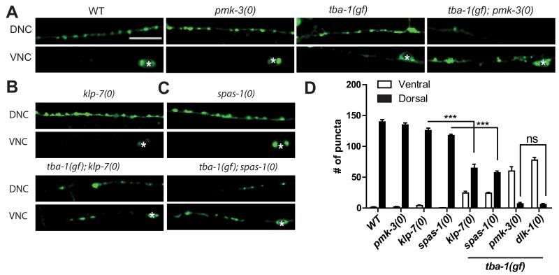
Figure 3. DLK-1 signals through PMK-3, and partly via KLP-7 and SPAS-1, in promoting DD remodeling.
(A-C) Images of DD neuron synapses (juIs137) in genotype as indicated. White asterisk mark DD cell bodies on the VNC. Scale bars: 10 μm.
(D) Quantification of ventral and dorsal DD synaptic puncta. Data are represented as mean ± SEM; n=10 animals per genotype. Statistics: One-Way ANOVA followed Tukey’s posttest; ***p<0.001, ns-not significant. See also Figure S2.

