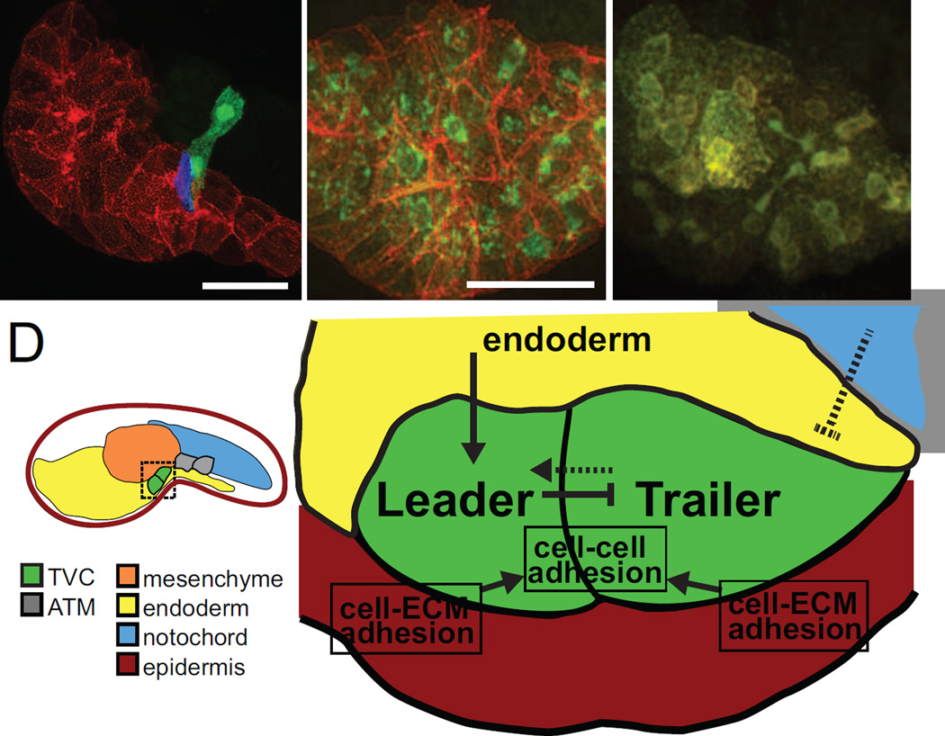
Figure 4. Surrounding tissues canalize TVCs towards directional migration.
(A) Initial tailbud embryo expressing membrane-localized reporter hCD4::mCherry in the endoderm (red), and GFP in the B7.5 lineage (green). Leader and trailer TVCs are shown. Surface contact analysis between TVC and endoderm indicates that only the presumptive leader TVC contacts the endoderm (blue). Scale bar, 40 µm. (B) Localization of membrane localized protein hCD4::mCherry (red) at cell membranes and KDEL receptor KDELR::GFP (green) labelling the endoplasmic reticulum (ER) in control embryos. Scale bar, 40 µm. (C) In embryos expressing dnSar1, hCD4::mCherry accumulates in the ER and cannot be properly trafficked to the membrane, indicated by the co-localization of red and green at the ER, and the absence of red fluorescence at cell membranes. (D) Schematic mid-tailbud embryo showing TVCs (green) and ATMs (gray) and tissues analyzed by Gline et al. by tissue-specific disruption of the secretory pathway: epidermis (red), mesenchyme (orange), endoderm (yellow) and notochord (blue). Model for extrinsic tissue influences on TVCs and intercellular interactions during TVC migration. The endoderm (yellow) signals to the TVCs (green) to establish leader-trailer polarity and TVCs in turn signal to each other to maintain polarity during migration. The notochord (blue) possibly sends chemorepulsive signals to migrating TVCs. TVCs adhere to each other during migration and also to the underlying epidermis (red) through cell-ECM interactions.

