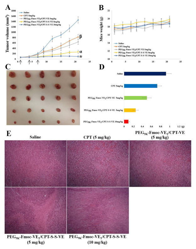
Fig. 7.
Antitumor efficacy of various CPT prodrug nanoformulations in 4T1.2 breast tumor model. Solid arrows indicate the i.v. injection. A: tumor volume. *p < 0.01, compared to PEG5K-Fmoc-VE2/CPT-S-S-VE (5 mg/kg); αp < 0.001, compared to PEG5K-Fmoc-VE2/CPT-VE (5 mg/kg) and CPT (5 mg/kg); βp < 0.001, compared to saline. B: mouse body weight. C: tumor images. D: tumor weight (g). E: Pathological examination of tumor tissues resected from the mice. Nuclei were stained by hematoxylin (blue), and extracellular matrix and cytoplasm were doped by eosin (red). Significant amount of necrotic/apoptotic cells were discerned in tumor tissues treated with PEG5K-Fmoc-VE2/CPT-S-S-VE over the other CPT formulations

