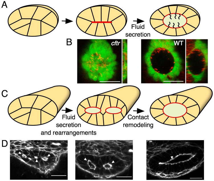
Figure 2. Fluid secretion during lumen formation.
(A) Schematic representation of Kupffer's vesicle (KV) lumen formation. The KV lumen forms from a group of initially disordered cells. Cellular rearrangements and fluid secretion generate a single central lumen with motile cilia that generate circular luminal fluid flow. (B) The importance of fluid secretion during lumen formation is illustrated by ventral and orthogonal views of the cftr mutant zebrafish KV, which contains a central plate of apical membrane and no luminal space, in contrast to the dramatic expansion of the WT organ (adapted from [15]). The KV epithelium (green) is labeled by sox17:GFP expression and the cilia are labeled by Arl13b (red). (C) In the zebrafish gut, lumen formation is initially characterized by lumen coalescence and expansion leading to a transient double lumen stage, which is resolved by Rab11-dependent contact remodeling. (D) This process is illustrated by stages of lumen formation in cross sections of the zebrafish gut (adapted from [27]). Filamentous actin (white) is marked by phalloidin.

