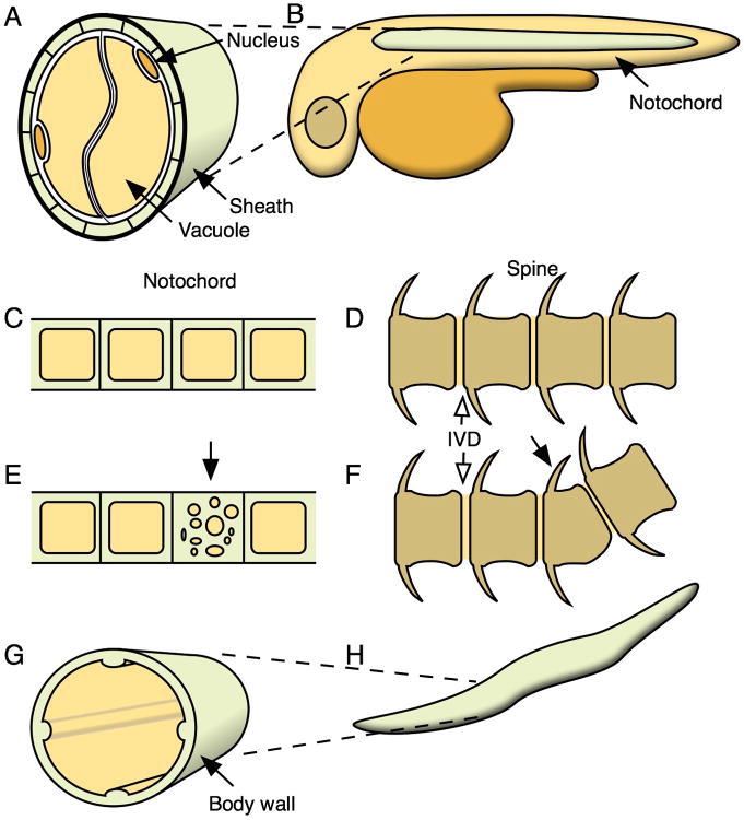
Figure 3. Fluid secretion shapes morphogenesis.
(A) The notochord is composed of cells containing large fluid filled vacuoles, surrounded by a sheath of extracellular matrix. (B) The hydrostatic pressure from the notochord acts as a hydrostatic skeleton and helps elongate the embryo. (C,D) A properly developed notochord serves as a scaffold for normal vertebral development. (E,F) Disruption of notochord vacuoles can lead to defects in spine morphogenesis and vertebral malformation. For simplicity, the sheath cells are not depicted in C and E. (G,H) The C. elegans body plan depends on internal fluid force restricted by extracellular matrix in the body wall.

